
Speaker
 Debraj Shome, MD, FRCS, FACS, MBA
The Esthetic Clinics, India
Dr. Debraj Shome is a top Facial Plastic Surgeon, Cosmetic Surgeon and Oculoplastic Surgeon. Dr. Shome co-founded The Esthetic Clinics. The Esthetic Clinics are a group of top-class centers, based in Mumbai, Hyderabad, Kolkata & New Delhi, India and dedicated to the disciplines of aesthetic surgery and skin care of the body. Dr. Shome is a Surgeon super specialized in Facial Plastic Surgery, Oculoplastic Surgery & Cosmetic Surgery & is a consultant at Breach Candy Hospital; Saifee Hospital, Girgaon, Mumbai; Apollo Spectra Hospital, Chembur, Mumbai & SL Raheja Fortis Hospital, Mahim, Mumbai, India. Dr. Shome also runs a NGO called Debabrata Auro Foundation which plays a role in the upliftment of the downtrodden. Dr. Shome is the ex-Head of the Institute of Aesthetic Surgery, Apollo Hospitals, Hyderabad, India. Dr. Shome was Visiting Faculty to the Department of Head & Neck Surgery at MD Anderson Cancer Center, Houston, USA.
Abstract
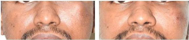
Introduction: Management of Vascular anomalies of the head and neck region using Intralesional bleomycin sclerotherapy has been done in the past. Various treatment options are available such as intralesional sclerotherapy, lasers, embolization and surgical excision. But a defined method of intervention is not yet established in the literature. This study aims to define a protocol for the management of extraoral head and neck vascular anomalies using intralesional bleomycin sclerotherapy.
Methods: A Prospective interventional study was carried out comprising of 10 patients aged between 18-65 years presenting with extra-oral vascular anomalies of head and neck region that have not been treated previously. Each vial containing 15 units of bleomycin solution (Bleocip; Cipla) was reconstituted with 2 ml of 0.9 % Normal Saline using BD 1 cc insulin syringe and a total of 30 Units reconstituted bleomycin solution was prepared. Reconstituted bleomycin solution was then injected 0.2 ml per site; 1 cm apart and simultaneous palpation of the regional vessels was performed. A total of 4 ml of 30 Units of bleomycin was injected intralesionally on each visit. A similar procedure was performed 4 weeks apart up to a minimum of 4 sessions for each patient. After 4 sessions, the reduction in the size was measured. Additional sessions were performed at multiple sites on the face when required. For the patients where the lesion was still present after 4 sessions, additional sessions were given. The maximum number of sessions for the study population were 8 sessions. The ice pack was applied for 15 minutes post procedure and changes such as bruising and local rise in temperature, pain, and pigmentation were observed. Post–operatively photographs were taken and also repeated during each visit.
Results: The mean age of patients was 38 years; presenting with Low flow Arteriovenous Malformation, lymphangioma, and hemangioma. A mean of 4 sessions were performed in all the patients. The reduction in size noted was approximately 45.83%. Additional 2 sessions were done in 3 patients and the reduction in the size of the lesion noted was 66.66%. In 1 patient, a total of 8 sessions were done and reduction in size was 85%. The mean reduction between 4th and 8th session was found to be 39.17% (p=0.0001). Side effects noted were bruising and pigmentation. No adverse complications or recurrence were encountered.
Conclusion: Intralesional bleomycin sclerotherapy provides a safe, outpatient and non-surgical method for the management of the extra-oral vascular anomalies of head and neck. This study provides a defined protocol which was not established till now. It gives a quantifiable approach with significant reduction in the size within a mean of 4 sessions. It also dissipates the need for surgical intervention in such patients and it works well in patients who have had a recurrence post-surgery as well.
Take Home Message
Intralesional bleomycin sclerotherapy provides a safe, outpatient, and non-surgical method for the management of the extra-oral vascular anomalies of the head and neck. This study provides a defined protocol that was not established till now.